
No hay productos en el carrito


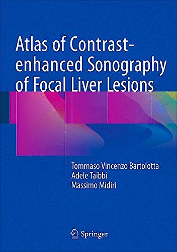
Atlas of Contrast-Enhanced Sonography of Focal Liver Lesions
Bartolotta, T. — Taibbi, A. — Midiri, M.
1ª Edición Agosto 2015
Inglés
Tapa dura
106 pags
200 gr
18 x 24 x null cm
ISBN 9783319175386
Editorial SPRINGER
LIBRO IMPRESO
-5%
103,99 €98,79 €IVA incluido
99,99 €94,99 €IVA no incluido
Recíbelo en un plazo de
2 - 3 semanas
Description
This book offers an image-based, comprehensive quick reference guide that will assist in the interpretation of contrast-enhanced ultrasound (CEUS) examinations of the liver in daily practice. It describes and depicts typical and atypical behavior of both common and less frequently observed focal liver lesions. For each type of lesion, the findings on pre- and post-contrast images are presented and key characteristics are highlighted. Individual chapters also focus on the assessment of response to locoregional and systemic treatment and the impact of European guidelines on CEUS. The Atlas of Contrast-Enhanced Sonography of Focal Liver Lesions will serve as an invaluable hands-on tool for practitioners who need to diagnose liver lesions using CEUS in the major clinical settings: oncology patients, cirrhotic patients, and patients with incidental focal liver lesions.
Contents
Authors
Tommaso Vincenzo Bartolotta, assistant Professor of radiology,
graduated from the University of Palermo, Italy, in 1993 and subsequently specialized
in Radiology, gaining his doctorate in Oncologic Radiology from the University
of Palermo in 2004. Since 2006 Head of the Ultrasound Unit in the department.
Prof. Bartolotta is the author of more than 300 scientific publications, including
original articles and book chapters. He is a board member of La Radiologia Medica
and World Journal of Radiology and is a reviewer for other journals. He has
been a member of the scientific organizing committees of numerous national and
international congresses and courses and an investigator or co-investigator
in many national and international research projects. His current research interests
include abdominal imaging (with a particular focus on study of the liver by
means of ultrasound, contrast-enhanced ultrasound, and magnetic resonance imaging),
thyroid imaging, and color Doppler ultrasound.
Adele Taibbi graduated from the University of Palermo, Italy, in 2002,
where she subsequently completed her specialization in Radiology and gained
her doctorate. Her current research interests include abdominal imaging, with
special focus on imaging of liver and gastrointestinal stromal tumors by means
of ultrasound, contrast-enhanced ultrasound, computed tomography, and magnetic
resonance imaging. Dr. Taibbi is author of more than 90 scientific publications,
including original articles and book chapters. She is a reviewer for European
Radiology and European Journal of Radiology and is an EPOS reviewer for the
European Congress of Radiology. She has been a speaker at more than 30 national
and international conferences.
Massimo Midiri is Full Professor of Radiology and Director of the Section
of Radiological Sciences, Department of Biopathology and Medical and Forensic
Biotechnologies, University Hospital “Paolo Giaccone”, Palermo,
Italy, which hosts a strong school of surgical oncology. In recent years, Dr.
Midiri has succeeded in establishing a new lab for preclinical studies at the
Section of Radiological Sciences, with installation of a very high magnetic
field strength (7T) MRI scanner for small animals. The Section of Radiological
Sciences also hosts the first and only Italian transcranial MRgFUS system for
the treatment of neurological disorders. Dr. Midiri is lead author or co-author
of more than 600 publications indexed on GoogleScholar, more than 250 of which
are indexed in PubMed.
© 2025 Axón Librería S.L.
2.149.0