
No hay productos en el carrito


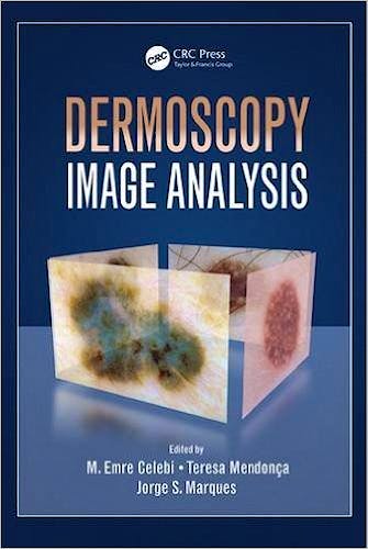
Dermoscopy Image Analysis
Celebi, M. — Mendonca, T. — Marques, J.
1ª Edición Octubre 2015
Inglés
Tapa dura
656 pags
1000 gr
15 x 23 x null cm
ISBN 9781482253269
Editorial CRC PRESS
LIBRO IMPRESO
-5%
227,15 €215,79 €IVA incluido
218,41 €207,49 €IVA no incluido
Recíbelo en un plazo de
2 - 3 semanas
Description
Demoscopy is a non-invasive skin imaging technique that uses optical magnification and either liquid immersion or cross- polarized lighting that makes subsurface structures more easily visible when compared to conventional clinical images. It allows the identification of dozens of morphological features, which is particularly important related to malignant melanoma. This book summarizes the state of the art for this exciting subfield within medical image analysis.
Features
- Provides complete coverage of dermoscopy image analysis, from preprocessing to classification
- Discusses state-of-the-art techniques and algorithms for dermoscopy image analysis
- Each chapter is authored by a leading expert in the field, from four different continents
- Contains numerous examples, illustrations, and tables summarizing results from quantitative studies
Table of Contents
Global Pattern Classification in Dermoscopic Images. Detection of Streaks in Dermoscopy Image Analysis. Towards a Robust Classification of Dermoscopy Images under Different Acquisition Setups. A State-of-the-Art Survey on Lesion Border Detection in Dermoscopy Images. Invariant Texture Descriptors for Dermoscopy. Colour in Dermoscopy Image Analysis: From Low-Level Features to High-Level Semantics. Accurate and Scalable System for Automatic Detection of Malignant Melanoma. Automatic Detection of Melanoma: An Application Based on the ABCD Criteria. Color Assessment for Melanoma Diagnosis Based on Perceptible Color Region. Streak-Detection in Dermoscopic Color Images using Localized Radial Flux of Principal Intensity Curvature. Where’s the Lesion? Variability in Human and Automated Segmentation of Dermatoscopically Imaged Melanocytic Lesions. Novel Segmentation-Classification Techniques for Dermoscopic Images Using Wavelet Transform. From Dermoscopy to Mobile Teledermatology. Computer Aided Diagnosis of Malignant Melanoma Based on Digital Dermoscopy. Comparison of Digital Signal Processing Techniques for Reticular Pattern Recognition in Melanoma Detection. Early Detection of Melanoma in Dermoscopy of Skin Lesion Images by Computer Vision Based System. PH2 - A Public Database for the Analysis of Dermoscopic Images.
© 2025 Axón Librería S.L.
2.149.0