
No hay productos en el carrito


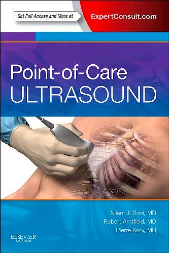
Point of Care Ultrasound
Arntfield, R. — Kory, P. — Soni, N.
1ª Edición Agosto 2014
Inglés
Tapa blanda
416 pags
900 gr
15 x 23 x 2 cm
ISBN 9781455775699
Editorial Saunders (W.B.) Co Ltd
Description
With portable, hand-carried ultrasound devices being more frequently implemented in medicine today, Point-of-Care Ultrasound will be a welcome resource for any physician or health care practitioner looking to further their knowledge and skills in point-of-care ultrasound. This comprehensive, portable handbook offers an easy-access format that combines electronic content and peer-reviewed videos to provide comprehensive, non-specialty-specific guidanceon this ever-evolving technology.
Features:
- Access all the facts with focused chapters covering a diverse range of topics, as well ascase-based examples that include ultrasound scans and videos.
- Understand the pearls and pitfalls of point-of-care ultrasound through contributions from experts at more than 30 institutions.
- View techniques more clearly than ever before. Illustrations and photos includetransducer position, cross-sectional anatomy, ultrasound cross sections, andultrasound images.
- Watch more than 200 ultrasound videos from real-life patients that demonstrate key findings. Available at Expert Consult, these videos are complemented by anatomical illustrations and text descriptions to maximize learning.
Table of Contents:
Section I: Fundamental Principles of Ultrasound
Chapter 1: Evolution of Point-of-Care Ultrasound
Chapter 2: Ultrasound Physics
Chapter 3: Transducers
Chapter 4: Orientation
Chapter 5: Basic Operation of an Ultrasound Machine
Chapter 6: Artifacts
Section II: Lung and Pleura
Chapter 7: Overview
Chapter 8: Lung and Pleural Ultrasound Technique
Chapter 9: Lung Ultrasound Interpretation
Chapter 10: Pleural Ultrasound Interpretation
Chapter 11: Lung and Pleural Procedures
Section III: Heart
Chapter 12: Overview
Chapter 13: Cardiac Ultrasound Technique
Chapter 14: Left Ventricular Function
Chapter 15: Right Ventricular Function
Chapter 16: Valves
Chapter 17: Pericardial Effusion
Chapter 18: Inferior Vena Cava
Section IV: Abdomen and Pelvis
Chapter 19: Gallbladder
Chapter 20: Kidneys
Chapter 21: Bladder
Chapter 22: Abdominal Aorta
Chapter 23: Abdominal Fluid
Chapter 24: First Trimester Pregnancy and Normal Female Reproductive System
Chapter 25: Testicular Ultrasound
Section V: Vascular System
Chapter 26: Lower Extremity Deep Venous Thrombosis
Chapter 27: Upper Extremity Deep Venous Thrombosis
Chapter 28: Central Venous Access
Chapter 29: Peripheral Venous Access
Chapter 30: Arterial Access
Section VI: Head and Neck
Chapter 31: Ocular Ultrasound
Chapter 32: Thyroid Gland
Chapter 33: Lymph Nodes
Section VII: Nervous System
Chapter 34: Peripheral Nerve Blocks
Chapter 35: Lumbar Puncture
Section VIII: Soft Tissues and Joints
Chapter 36: Skin and Soft Tissues
Chapter 37: Joints
Section IX: Clinical Scenarios and Protocols
Chapter 38: Dyspnea and Acute Respiratory Failure
Chapter 39: Abdominal Pain
Chapter 40: Hypotension and Shock
Chapter 41: Trauma
Chapter 42: Cardiac Arrest
Section X: Ultrasound Program Management
Chapter 43: Competence, Credentialing, and Certification
Chapter 44: Equipment, Image Archiving, and Billing
Index
By Nilam J Soni, MD, Associate Professor of Medicine, Division of Hospital Medicine, University of Texas Health Science Center San Antonio, San Antonio, Texas, USA ; Robert Arntfield, MD, Assistant Professor of Medicine, Divisions of Emergency Medicine and Critical Care Medicine, Western University, London, Ontario, Canada and Pierre Kory, MPA, MD, Associate Professor of Medicine, Division of Pulmonary & Critical Care Medicine, Icahn School of Medicine at Mount Sinai, Beth Israel Medical Center, New York, New York, USA
© 2026 Axón Librería S.L.
2.149.0