
No hay productos en el carrito


Fundamentals of Oral and Maxillofacial Radiology
Hubar, J.
1ª Edición Julio 2017
Inglés
Tapa blanda
264 pags
500 gr
17 x 24 x null cm
ISBN 9781119122210
Editorial WILEY
LIBRO IMPRESO
-5%
110,95 €105,40 €IVA incluido
106,68 €101,35 €IVA no incluido
Recíbelo en un plazo de
2 - 3 semanas
LIBRO ELECTRÓNICO
-5%
93,87 €89,18 €IVA incluido
90,26 €85,75 €IVA no incluido
Acceso On Line
Inmediato
Description
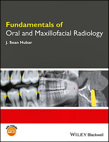
Fundamentals of Oral and Maxillofacial Radiology provides a concise overview of the principles of dental radiology, emphasizing their application to clinical practice.
- Distills foundational knowledge on oral radiology in an accessible guide
- Uses a succinct, easy-to-follow approach
- Focuses on practical applications for radiology information and techniques
- Presents summaries of the most common osseous pathologic lesions and dental anomalies
- Includes companion website with figures from the book in PowerPoint and x-ray puzzles
Table of Contents
Acknowledgments
Section One: FUNDAMENTALS
A. Introduction
B. History
C. Generation of x rays
D. Exposure controls
E. Radiation dosimetry
F. Radiation biology
G. Radiation protection
H. Patient selection criteria
I. Analog film versus digital image
J. What do dental x-ray images reveal?
K. Intraoral imaging techniques
L. Intraoral technique errors
M. Extraoral imaging techniques
N. Quality assurance
O. Infection control
P. Occupational radiation exposure monitoring
Q. Hand-held x-ray systems
Section Two: INTERPRETATION
R. Localization of objects (SLOB rule)
S. Recommendations for interpreting images
T. X-ray puzzles: spot the differences
U. Radiographic anatomy
V. Dental caries
W. Dental anomalies
X. Osseous pathology (alphabetic)
Y. Lagniappe (miscellaneous oddities)
Section Three: APPENDICES
Appendix 1: FDA recommendations for prescribing dental x-ray images
Appendix 2: X-radiation concerns of patients: question and answer format
Appendix 3: Helpful tips for difficult patients
Appendix 4: Deficiencies of dental imaging terminology
Appendix 5: Tools for differential diagnosis
Appendix 6: Table of radiation units
Appendix 7: Table of anatomic landmarks
Appendix 8: Table of dental anomalies
Appendix 9: Table of osseous pathology
Appendix 10: Common abbreviations and acronyms
Appendix 11: Glossary of terms
References
Index
Author Information
J. Sean Hubar, DMD, MS, is Professor in the Department of Diagnostic Sciences at Louisiana State University School of Dentistry in New Orleans, Louisiana, USA.
© 2025 Axón Librería S.L.
2.149.0