
No hay productos en el carrito


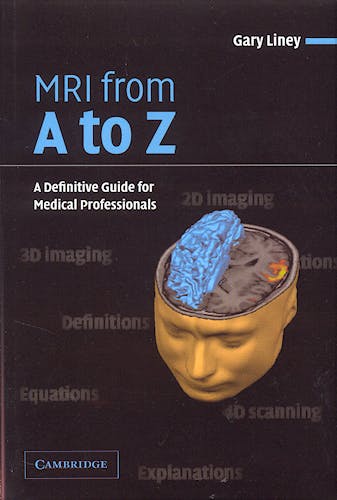
Mri from a to Z. a Definitive Guide for Medical Professionals
Liney, G.
1ª Edición Marzo 2005
Inglés
Tapa blanda
270 pags
292 gr
12 x 14 x 1 cm
ISBN 9780521606387
Editorial CAMBRIDGE
LIBRO IMPRESO
-5%
64,17 €60,96 €IVA incluido
61,70 €58,62 €IVA no incluido
Recíbelo en un plazo de
2 - 3 semanas
From ‘AB systems’ to ‘Zipper artefact’ - even for the experienced practitioner in MRI, the plethora of technical terms and acronyms can be daunting and bewildering. This concise but comprehensive guide provides an effective and practical introduction to the full range of this terminology. It will be an invaluable source of reference for all students, trainees and medical professionals working with MRI. More than 800 terms commonly encountered in MR Imaging and Spectroscopy are clearly defined, explained and cross-referenced. Illustrations are used to enhance and explain many of the definitions, and references to further reading will point the reader to more in-depth coverage. As well as being a compendium of terms from A to Z, the volume concludes with a useful collection of appendices, which tabulate many of the key constants, properties and equations of relevance.
- Concise, clear and comprehensive coverage of MRI terminology
- Definitions are enhanced with illustrations and references to further reading
- Appendices provide a core listing of key data, constants and equations
- Main glossary
Aa
AB systems
Referring to molecules exhibiting multiply split MRS peaks due to spin-spin interactions. In an AB system, the chemical shift between the spins is of similar magnitude to the splitting constant ( J ). A common example is citrate (abundant in the normal prostate). Citrate consists of two pairs of methylene protons (A and B, see Appendix VI) that are strongly coupled such that:
where ?A, ?B are the resonating frequencies of the two protons. A tall central doublet is split into two smaller peaks either side, which are not usually resolved in vivo at 1.5 tesla. Citrate exhibits strong echo modulation.
See also J-coupling and AX systems.
Reference R. B. Mulkern & J. L. Bowers (1994). Density matrix calculations of AB spectra from multipulse sequences: quantum mechanics meets spectroscopy. Concepts Magn. Reson. 6, 1–23.
Absolute peak area quantification
MR spectroscopy method of using peak area ratios where the denominator is the water peak. The areas are adjusted for differences in relaxation times, and the actual concentration of the metabolite is determined from:
where [w] is the concentration of water and S0 are the peak area amplitudes of the metabolite and water signals at equilibrium, i.e. having been corrected for relaxation, which has occurred at the finite time of measurement. The factor 2/n corrects for the number of protons contributing to the signal (here 2 is for water).
Note: [w] is taken as 55.55 Mol/kg.
Reference P. B. Barker, B. J. Soher, S. J. Blackband, J. C. Chatham, V. P. Mathews & R. N. Bryan (1993). Quantitation of proton NMR spectra of the human brain using tissue water as an internal concentration reference. NMR Biomed. 6, 89–94.
Acoustic noise
The audible noise produced by the scanner. Caused by vibrations in the gradient coils induced by the rapidly oscillating currents passing through them in the presence of the main magnetic field. Ear protection must by worn by patients because of this noise. Gradient-intensive sequences, e.g. 3-D GRE, EPI, produce the highest noise levels. Typically, the recorded noise level may be weighted (dB (A) scale) to account for the frequency response of the human ear. Values of 115 dB (A) have been recorded with EPI. The Lorentz force, and therefore noise level, increases with field strength (typically a 6 dB increase from 1.5 to 3.0 tesla). Current methods to combat noise include mounting the gradient coils to the floor to reduce vibrations and lining the bore with a vacuum. More sophisticated measures include active noise reduction.
See also bore liner and vacuum bore.
Reference F. G. Shellock, M. Ziarati, D. Atkinson & D. Y. Chen (1998). Determination of gradient magnetic field-induced acoustic noise associated with the use of echoplanar and three-dimensional fast spin echo techniques. J. Magn. Reson. Imag. 8, 1154.
Acquisition time
Time taken to acquire an MR image. For a spin-echo sequence it is given by:
Np × NA × TR
where Np is the number of phase encoding steps, NA is the number of signal averages, and TR is the repetition time. Shorter scan times means a trade-off in image quality in terms of resolution (Np), SNR (NA) and contrast (TR). Scan times may also be reduced by using parallel imaging.
In gradient-echo sequences with very short TR times, the above equation includes a factor for the number of slices acquired............
© 2025 Axón Librería S.L.
2.149.0