
No hay productos en el carrito


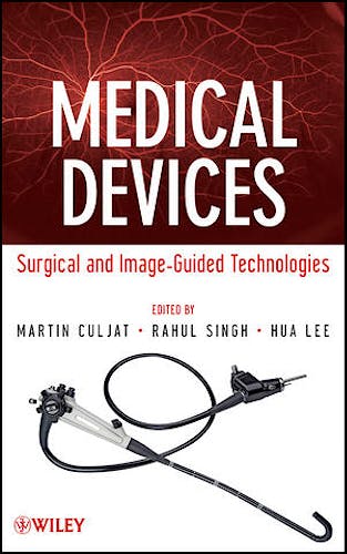
Medical Devices: Surgical and Image-Guided Technologies
Culjat, M. — Singh, R. — Lee, H.
1ª Edición Noviembre 2012
Inglés
Tapa dura
456 pags
747 gr
16 x 24 x 3 cm
ISBN 9780470549186
Editorial JOHN WILEY & SONS
LIBRO IMPRESO
-5%
162,56 €154,43 €IVA incluido
156,31 €148,49 €IVA no incluido
Recíbelo en un plazo de
2 - 3 semanas
LIBRO ELECTRÓNICO
-5%
144,29 €137,08 €IVA incluido
138,74 €131,81 €IVA no incluido
Acceso On Line
Inmediato
Description
Medical Devices is a textbook for an introductory seminar course on biomedical devices and technology. The book covers devices and systems in diagnostic, surgical, and implant procedures, prepared by the much-respected faculty members at the UCLA School of Medicine. Technical contents are presented in a comprehensive manner, consistent with first-year students’ background level in mathematics, physics, chemistry, and biology. The chapters are written and organized in the form of independent modules, such that lectures can be configured with a high degree of flexibility from year to year.To gauge a preliminary assessment of the effectiveness of this book's technical coverage, nine of the authors participated in a one-quarter seminar course at UC Santa Barbara, receiving superb ratings and reviews. The class attracted students from all engineering majors, as well as the pre-med program, with a breadth of audience and interest level that this book carries through gracefully.
Table of contents
PREFACE
CONTRIBUTORS
PART I INTRODUCTION TO MEDICAL DEVICES 1
1. Introduction 3
Martin Culjat
- 1.1 History of Medical Devices 3
- 1.2 Medical Device Terminology 6
- 1.3 Purpose of the Book 10
2. Design of Medical Devices 11
Gregory Nighswonger
- 2.1 Introduction 11
- 2.2 The Medical Device Design Environment 11
- 2.2.1 US Regulation 12
- 2.2.2 Differences in European Regulation 13
- 2.2.3 Standards 14
- 2.3 Basic Design Phases 15
- 2.3.1 Feasibility 15
- 2.3.2 Planning and Organization—Assembling the Design Team 16
- 2.3.3 When to Involve Regulatory Affairs 17
- 2.3.4 Conceptualizing and Review 17
- 2.3.5 Testing and Refinement 20
- 2.3.6 Proving the Concept 20
- 2.3.7 Pilot Testing and Release to Manufacturing 22
- 2.4 Postmarket Activities 25
- 2.5 Final Note 25
PART II MINIMALLY INVASIVE DEVICES AND TECHNIQUES 27
3. Instrumentation for Laparoscopic Surgery 29
Camellia Racu-Keefer, Scott Um, Martin Culjat, and Erik Dutson
- 3.1 Introduction 29
- 3.2 Basic Principles 31
- 3.3 Laparoscopic Instrumentation 34
- 3.3.1 Trocars 34
- 3.3.2 Standard Laparoscopic Instruments 37
- 3.3.3 Additional Laparoscopic Instruments 42
- 3.3.4 Specimen Retrieval Bags 44
- 3.3.5 Disposable Instruments 44
- 3.4 Innovative Applications 45
- 3.5 Summary and Future Applications 46
4. Surgical Instruments in Ophthalmology 49
Allen Y. Hu, Robert M. Beardsley, and Jean-Pierre Hubschman
- 4.1 Introduction 49
- 4.2 Cataract Surgery 51
- 4.2.1 Basic Technique 51
- 4.2.2 Principles of Phacoemulsification 52
- 4.2.3 Phacoemulsification Instruments 54
- 4.2.4 Phacoemulsification Systems 55
- 4.2.5 Future Directions 56
- 4.3 Vitreoretinal Surgery 56
- 4.3.1 Basic Techniques 56
- 4.3.2 Principles of Vitrectomy 57
- 4.3.3 Vitrectomy Instruments 58
- 4.3.4 Vitrectomy Systems 60
- 4.3.5 Future Directions 60
- 4.4 Other Ophthalmic Surgical Procedures 61
- 4.5 Conclusion 62
5. Surgical Robotics 63
Jacob Rosen
- 5.1 Introduction 63
- 5.2 Background and Leading Concepts 63
- 5.2.1 Human–Machine Interfaces: System Approach 65
- 5.2.2 Tissue Biomechanics 70
- 5.2.3 Teleoperation 72
- 5.2.4 Image-Guided Surgery 78
- 5.2.5 Objective Assessment of Skill 79
- 5.3 Commercial Systems 80
- 5.3.1 ROBODOC (Curexo Technology Corporation) 80
- 5.3.2 daVinci (Intuitive Surgical) 83
- 5.3.3 Sensei X (Hansen Medical) 84
- 5.3.4 RIO MAKOplasty (MAKO Surgical Corporation) 86
- 5.3.5 CyberKnife (Accuray) 89
- 5.3.6 Renaissance™ (Mazor Robotics) 91
- 5.3.7 ARTAS System (Restoration Robotics, Inc.) 92
- 5.4 Trends and Future Directions 93
6. Catheters in Vascular Therapy 99
Axel Boese
- 6.1 Introduction 99
- 6.2 Historic Overview 100
- 6.3 Catheter Interventions 102
- 6.4 Catheter and Guide Wire Shapes and Configurations 105
- 6.4.1 Catheters 105
- 6.4.2 Guide Wires 113
- 6.5 Conclusion 116
PART III ENERGY DELIVERY DEVICES AND SYSTEMS 119
7. Energy-Based Hemostatic Surgical Devices 121
Amit P. Mulgaonkar, Warren Grundfest, and Rahul Singh
- 7.1 Introduction 121
- 7.2 History of Energy-Based Hemostasis 122
- 7.3 Energy-Based Surgical Methods and Their Effects on Tissues 125
- 7.3.1 Disambiguation 126
- 7.3.2 Thermal Effects on Tissues 127
- 7.4 Electrosurgery 128
- 7.4.1 Electrosurgical Theory 128
- 7.4.2 Cutting and Coagulation Techniques 130
- 7.4.3 Equipment 131
- 7.4.4 Considerations and Complications 133
- 7.5 Future Of Electrosurgery 134
- 7.6 Conclusion 135
8. Tissue Ablation Systems 137
Michael Douek, Justin McWilliams, and David Lu
- 8.1 Introduction 137
- 8.2 Evolving Paradigms in Cancer Therapy 138
- 8.3 Basic Ablation Categories and Nomenclature 140
- 8.4 Hyperthermic Ablation 140
- 8.5 Fundamentals of In Vivo Energy Deposition 141
- 8.6 Hyperthermic Ablation: Optimizing Tissue Ablation 143
- 8.7 Radiofrequency Ablation 144
- 8.8 RFA: Basic Principles 145
- 8.9 RFA: In Vivo Energy Deposition 145
- 8.10 Optimizing RFA 147
- 8.11 Other Hyperthermic Ablation Techniques 149
- 8.11.1 Microwave Ablation (MWA) 149
- 8.11.2 MWA: Basic Principles 149
- 8.11.3 MWA: In Vivo Energy Deposition 151
- 8.11.4 Optimizing MWA 152
- 8.12 Laser Ablation 153
- 8.13 Hypothermic Ablation 154
- 8.13.1 Cryoablation: Basic Concepts 154
- 8.13.2 Cryoablation: In Vivo Considerations 154
- 8.13.3 Optimizing Cryoablation Systems 154
- 8.14 Chemical Ablation 157
- 8.15 Novel Techniques 158
- 8.15.1 High Intensity Focused Ultrasound (HIFU) 158
- 8.15.2 Irreversible Electroporation (IRE) 159
- 8.16 Tumor Ablation and Beyond 160
9. Lasers in Medicine 163
Zachary Taylor, Asael Papour, Oscar Stafsudd, and Warren Grundfest
- 9.1 Introduction 163
- 9.1.1 Historical Perspective 164
- 9.1.2 Basic Operational Concepts 165
- 9.1.3 First Experimental MASER (Microwave Amplification by Stimulated Emission of Radiation) 166
- 9.2 Laser Fundamentals 167
- 9.2.1 Two-Level Systems and Population Inversion 167
- 9.2.2 Multiple Energy Levels 167
- 9.2.3 Mode of Operation 169
- 9.2.4 Beams and Optics 171
- 9.3 Laser Light Compared to Other Sources of Light 174
- 9.3.1 Temporal Coherence 174
- 9.3.2 Spectral Coherence (Line Width) 175
- 9.3.3 Beam Collimation 177
- 9.3.4 Short Pulse Duration 177
- 9.3.5 Summary 178
- 9.4 Laser–Tissue Interactions 178
- 9.4.1 Biostimulation 178
- 9.4.2 Photochemical Interactions 179
- 9.4.3 Photothermal Interactions 180
- 9.4.4 Ablation 180
- 9.4.5 Photodisruption 181
- 9.5 Lasers in Diagnostics 181
- 9.5.1 Optical Coherence Tomography 181
- 9.5.2 Fluorescence Angiography 184
- 9.5.3 Near Infrared Spectroscopy 185
- 9.6 Laser Treatments and Therapy 186
- 9.6.1 Overview of Current Medical Applications of Laser Technology 186
- 9.6.2 Retinal Photodynamic Therapy (Photochemical) 188
- 9.6.3 Transpupillary Thermal Therapy (TTT) (Photothermal) 188
- 9.6.4 Vascular Birth Marks (Photocoagulation) 190
- 9.6.5 Laser Assisted Corneal Refractive Surgery (Ablation) 191
- 9.7 Conclusions 196
PART IV IMPLANTABLE DEVICES AND SYSTEMS 197
10. Vascular and Cardiovascular Devices 199
Dan Levi, Allan Tulloch, John Ho, Colin Kealey, and David Rigberg
- 10.1 Introduction 199
- 10.2 Biocompatibility Considerations 200
- 10.3 Materials 202
- 10.3.1 316L Stainless Steel 203
- 10.3.2 Nitinol 203
- 10.3.3 Cobalt–Chromium Alloys 204
- 10.4 Stents 204
- 10.5 Closure Devices 206
- 10.6 Transcatheter Heart Valves 208
- 10.7 Inferior Vena Cava Filters 212
- 10.8 Future Directions–Thin Film Nitinol 214
- 10.9 Conclusion 216
11. Mechanical Circulatory Support Devices 219
Colin Kealey, Paymon Rahgozar, and Murray Kwon
- 11.1 Introduction 219
- 11.2 History 220
- 11.3 Basic Principles 221
- 11.3.1 Biocompatibility and Mechanical Circulatory Support Devices 221
- 11.3.2 Hemocompatibility: Microscopic Considerations 222
- 11.3.3 Hemocompatibility: Macroscopic Considerations 223
- 11.4 Engineering Considerations in Mechanical Circulatory Support 223
- 11.4.1 Overview 223
- 11.4.2 Pump Design 225
- 11.4.3 Positive Displacement Pumps 225
- 11.4.4 Rotary Pumps 226
- 11.4.5 Pulsatile Versus Nonpulsatile Flow 228
- 11.5 Devices 228
- 11.5.1 The HeartMate XVE Left Ventricular Assist System 228
- 11.5.2 The HeartMate II Left Ventricular Assist System 231
- 11.5.3 Short-Term Mechanical Circulatory Support: The Intraaortic Balloon Pump 234
- 11.5.4 Pediatric Mechanical Circulatory Support: The Berlin Heart 237
- 11.6 The Future of MCS Devices 239
- 11.6.1 CorAide 239
- 11.6.2 HeartMate III 239
- 11.6.3 HeartWare 240
- 11.6.4 VentrAssist 240
- 11.7 Summary 240
12. Orthopedic Implants 241
Sophia N. Sangiorgio, Todd S. Johnson, Jon Moseley, G. Bryan Cornwall, and Edward
Ebramzadeh
- 12.1 Introduction 241
- 12.1.1 Overview 241
- 12.1.2 History 243
- 12.2 Basic Principles 244
- 12.2.1 Optimization for Strength and Stiffness 245
- 12.2.2 Maximization of Implant Fixation to Host Bone 250
- 12.2.3 Minimization of Degradation 251
- 12.2.4 Sterilization of Implants and Instrumentation 253
- 12.3 Implant Technologies 253
- 12.3.1 Total Hip Replacement 254
- 12.3.2 Technology in Total Knee Replacement 263
- 12.3.3 Technology in Spine Surgery 268
- 12.4 Summary 272
PART V IMAGING AND IMAGE-GUIDED TECHNIQUES 275
13. Endoscopy 277
Gregory Nighswonger
- 13.1 Introduction 277
- 13.2 Ancient Origins 278
- 13.3 Modern Endoscopy 280
- 13.3.1 Creating Cold Light 280
- 13.3.2 Introduction of Rod-Lens Technology 280
- 13.4 Principles of Modern Endoscopy 283
- 13.4.1 Optics 284
- 13.4.2 Mechanics 284
- 13.4.3 Electronics 284
- 13.4.4 Software 285
- 13.5 The Imaging Chain 285
- 13.5.1 Light Source (1) 286
- 13.5.2 Telescope (2) 286
- 13.5.3 Camera Head (3) 287
- 13.5.4 Camera CCU (4) 287
- 13.5.5 Video Cables (5) 287
- 13.5.6 Monitor (6) 287
- 13.5.7 Image Management Systems (7) 288
- 13.6 Endoscopes for Today 288
- 13.6.1 Rigid Endoscopes—Designs to Enhance Functionality 289
- 13.6.2 Less Traumatic Ureterorenoscopes 290
- 13.6.3 Advances in Flexible Endoscope Design 291
- 13.6.4 Broader Functionality with New Technologies 294
- 13.6.5 Enhancing Video Capabilities 299
- 13.7 Endoscopy’s Future 301
14. Medical Ultrasound Devices 303
Rahul Singh and Martin Culjat
- 14.1 Introduction 303
- 14.2 Basic Principles of Ultrasound 304
- 14.2.1 Basic Acoustic Physics 304
- 14.2.2 Reflection and Refraction 307
- 14.2.3 Attenuation 307
- 14.2.4 Piezoelectricity 308
- 14.2.5 Ultrasound Systems 310
- 14.2.6 Resolution and Bandwidth 312
- 14.2.7 Beam Characteristics 314
- 14.3 Ultrasound Transducer Design 316
- 14.3.1 Piezoelectric Material 317
- 14.3.2 Backing Layers and Damping 318
- 14.3.3 Matching Layers 318
- 14.3.4 Mechanical Focusing 319
- 14.3.5 Electrical Matching 320
- 14.3.6 Sector Scanners 320
- 14.3.7 Array Transducers 322
- 14.3.8 Transducer Array Fabrication 325
- 14.3.9 Regulatory Considerations 327
- 14.4 Applications of Medical Ultrasound 329
- 14.4.1 Image Guidance Applications 330
- 14.4.2 Intravascular and Intracardiac Applications 332
- 14.4.3 Intraoral and Endocavity Applications 333
- 14.4.4 Surgical Applications 334
- 14.4.5 Ophthalmic Ultrasound 335
- 14.4.6 Doppler and Doppler Applications 336
- 14.4.7 Therapeutic Applications 336
- 14.5 The Future of Medical Ultrasound 338
15. Medical X-ray Imaging 341
Mark Roden
- 15.1 Introduction 341
- 15.2 X-ray Physics 342
- 15.2.1 Photon Interactions with Matter 342
- 15.2.2 Clinical Production of X-rays 343
- 15.2.3 Patient Dose Considerations 346
- 15.3 Two-Dimensional Image Acquisition 348
- 15.4 Image Acquisition Technologies and Techniques 351
- 15.4.1 Film 351
- 15.4.2 Computed Radiography 354
- 15.4.3 Digital Radiography 358
- 15.4.4 Clinical Applications of 2D X-ray Techniques 360
- 15.5 Basic 2D Processing Techniques 361
- 15.5.1 Independent Pixel Operations 362
- 15.5.2 Grouped Pixel Operations 363
- 15.5.3 Image Transformation Operations 366
- 15.6 Real-Time X-ray Imaging 367
- 15.6.1 Fluoroscopy Technology 367
- 15.6.2 Angiography 370
- 15.7 Three-Dimensional X-ray Imaging 372
- 15.8 Conclusion 373
16. Navigation in Neurosurgery 375
Jean-Jacques Lemaire, Eric J. Behnke, Andrew J. Frew, and Antonio A. F. DeSalles
- 16.1 Basics of Neurosurgery 375
- 16.1.1 General Technical Issues in Neurosurgery 375
- 16.1.2 Instrumentation in Neurosurgery 376
- 16.1.3 Complications 377
- 16.1.4 Functional Neurosurgery 378
- 16.1.5 Stereotactic Neurosurgery 378
- 16.1.6 Neuroimaging for Neurosurgery 379
- 16.2 Introduction to Neuronavigation 381
- 16.3 Neuronavigation Systems 381
- 16.3.1 The Tracking System 382
- 16.3.2 The Display Unit 383
- 16.3.3 The Control Unit 385
- 16.4 Implementation of Neuronavigation 386
- 16.4.1 Surgical Planning 386
- 16.4.2 Patient Registration 387
- 16.4.3 Navigation 389
- 16.5 Augmented Reality and Virtual Reality 390
- 16.6 Summary/Future 391
REFERENCES 395
INDEX 425
© 2026 Axón Librería S.L.
2.149.0