
No hay productos en el carrito


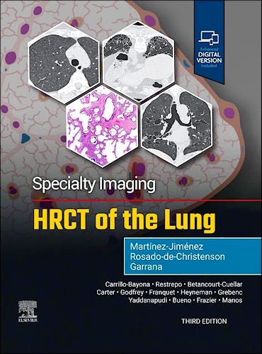
Specialty Imaging: HRCT of the Lung
Martínez-Jiménez, S. — Rosado-De-Christenson, M. — Garrana, S.
3ª Edición Julio 2025
Inglés
Tapa dura
632 pags
2000 gr
22 x 28 x 3 cm
ISBN 9780443286360
Editorial ELSEVIER
LIBRO IMPRESO
-5%
265,95 €252,65 €IVA incluido
255,72 €242,93 €IVA no incluido
Recíbelo en un plazo de
2 - 3 semanas
- SECTION 1: FUNDAMENTALS OF HRCT
- Overview of HRCT
- Approach to HRCT Interpretation
- ANATOMY
- Interstitial Compartments
- Secondary Pulmonary Lobule
- Gravitational Changes (Dependent Atelectasis)
- Age-Related Changes
- Normal Inspiration and Expiration
- TERMINOLOGY AND SIGNS
- 3-Density Sign (Formerly Head Cheese Sign)
- Airspace Nodules
- Anterior Upper Lobe Sign
- Crazy Paving
- Cystic Lung Disease
- Exuberant Honeycombing
- Finger-in-Glove Sign
- Flame-Shaped Nodules
- Four Corners Sign
- Ground Glass
- Halo Sign
- Honeycombing (Peribronchovascular)
- Honeycombing (Subpleural)
- Honeycombing (Unilateral)
- Micronodules
- Mosaic Attenuation and Air-Trapping
- Reversed Halo Sign
- Signet-Ring Sign
- Straight-Edge Sign
- Tree-in-Bud Opacities
- Thick-Walled Cystic Lesions (Stalactite and Stalagmite Sign)
- DISTRIBUTION
- Peribronchovascular
- Centrilobular
- Perilymphatic
- Random
- Peripheral
- SECTION 2: PATHOLOGIC PATTERNS OF INJURY
- Approach to Pathologic Patterns of Injury
- Diffuse Alveolar Damage
- Diffuse Alveolar Hemorrhage With Capillaritis
- Organizing Pneumonia
- Constrictive Bronchiolitis
- SECTION 3: LARGE AIRWAYS DISEASE
- Approach to Large Airways Disease
- Bronchiectasis
- Allergic Bronchopulmonary Aspergillosis
- Williams-Campbell Syndrome
- Mounier-Kuhn Syndrome
- Bronchocentric Granulomatosis
- SECTION 4: SMALL AIRWAYS DISEASE
- Approach to Small Airways Disease
- Infectious Bronchiolitis
- Asthma
- Diffuse Aspiration Bronchiolitis
- Respiratory Bronchiolitis
- Follicular Bronchiolitis
- Hypersensitivity Pneumonitis
- Diffuse Panbronchiolitis
- Idiopathic Constrictive Bronchiolitis
- Swyer-James-MacLeod Syndrome
- Bronchiolitis Obliterans Syndrome
- Diffuse Idiopathic Pulmonary Neuroendocrine Cell Hyperplasia (DIPNECH)
- SECTION 5: INFECTION
- Approach to Infection
- Bacterial Pneumonia
- Mycoplasma Pneumonia
- Staphylococcus aureus Pneumonia
- Viral Pneumonia
- Influenza
- COVID-19
- Respiratory Syncytial Virus
- Cytomegalovirus
- Tuberculosis
- Nontuberculous Mycobacterial Infection
- Endemic Fungal Infection
- Cryptococcosis
- Aspergillosis
- Pneumocystis Pneumonia
- Candidiasis
- SECTION 6: PNEUMOCONIOSIS
- Approach to Pneumoconiosis
- Silicosis and Coal Worker's Pneumoconiosis
- Asbestosis
- Berylliosis
- Talcosis
- Hard Metal Lung Disease
- SECTION 7: NEOPLASMS
- Approach to Neoplasms
- Invasive Mucinous Adenocarcinoma (Diffuse)
- Diffuse and Multifocal Lung Cancer
- Lymphangitic Carcinomatosis
- Hematogenous Metastases
- Endovascular Metastases and Tumor Emboli
- Kaposi Sarcoma
- Lymphangioleiomyomatosis
- Reactive Lymphoproliferative Disorders
- Neoplastic Lymphoproliferative Disorders
- Pulmonary Langerhans Cell Histiocytosis
- Erdheim-Chester Disease
- Rosai-Dorfman Disease
- Diffuse Pulmonary Meningotheliomatosis (DPM)
- SECTION 8: INTERSTITIAL PNEUMONIAS
- Approach to Interstitial Pneumonias
- Interstitial Lung Abnormality (ILA)
- Idiopathic Pulmonary Fibrosis
- Progressive Pulmonary Fibrosis (PPF)
- Idiopathic Nonspecific Interstitial Pneumonia
- Cryptogenic Organizing Pneumonia
- Acute Exacerbation of Interstitial Lung Disease
- Acute Interstitial Pneumonia
- Idiopathic Lymphoid Interstitial Pneumonia
- Pleuroparenchymal Fibroelastosis
- Airway-Centered Interstitial Fibrosis
- Interstitial Pneumonia With Autoimmune Features (IPAF)
- Approach to Smoking-Related Interstitial Lung Diseases
- Respiratory Bronchiolitis-Interstitial Lung Disease
- Desquamative Interstitial Pneumonia
- Syndrome of Combined Pulmonary Fibrosis and Emphysema
- Smoking-Related Interstitial Fibrosis (SRIF)
- Airspace Enlargement With Fibrosis (AEF)
- SECTION 9: AUTOIMMUNE DISEASES
- Approach to Connective Tissue Disease-Associated Interstitial Lung Disease
- Rheumatoid Arthritis
- Progressive Systemic Sclerosis
- Idiopathic Inflammatory Myopathies
- Sjögren Syndrome
- Mixed Connective Tissue Disease
- Systemic Lupus Erythematosus
- Granulomatosis With Polyangiitis (GPA)
- Eosinophilic Granulomatosis With Polyangiitis
- Microscopic Polyangiitis
- Ankylosing Spondylitis
- Inflammatory Bowel Disease-Associated Lung Disease
- SECTION 10: VASCULAR DISEASE
- Approach to Vascular Disease
- Pulmonary Edema
- Hepatopulmonary Syndrome
- Pulmonary Hypertension
- Pulmonary Venoocclusive Disease/Pulmonary Capillary Hemangiomatosis
- Excipient Lung Disease (Talc/Cellulose Granulomatosis)
- Chronic Thromboembolic Pulmonary Hypertension
- SECTION 11: INHALATIONAL, INFLAMMATORY, METABOLIC, AND POST TREATMENT
- ASPIRATION/INHALATION
- Spectrum of Aspiration-Related Disorders
- Lipoid Pneumonia
- Inhalational Lung Injury
- E-Cigarette or Vaping Product Use-Associated Lung Injury (EVALI)
- Recurrent Gastric Acid Aspiration and Dendriform Pulmonary Ossification
- INFLAMMATORY
- Sarcoidosis
- Acute Eosinophilic Pneumonia
- Chronic Eosinophilic Pneumonia
- Other Eosinophilic Disorders
- METABOLIC OR DEGENERATIVE
- Amyloidosis
- Light Chain Deposition Disease
- Pulmonary Alveolar Proteinosis
- Metastatic Pulmonary Calcification
- Diffuse Pulmonary Ossification
- Emphysema
- Idiopathic Pulmonary Hemosiderosis
- POST TREATMENT
- Radiation-Induced Lung Disease
- Drug-Induced Lung Disease
- Immune Checkpoint Inhibitor-Related Pneumonitis
- ASPIRATION/INHALATION
- SECTION 12: CONGENITAL
- Approach to Congenital
- Familial Idiopathic Pulmonary Fibrosis
- Birt-Hogg-Dubé Syndrome
- Hermansky-Pudlak Syndrome
- Tuberous Sclerosis Complex
- Neurofibromatosis
- Alveolar Microlithiasis
- α-1 Antitrypsin Deficiency
- Primary Ciliary Dyskinesia
- Chronic Granulomatous Disease
- Common Variable Immunodeficiency
- Primary Immunodeficiency Disorders
- Cystic Fibrosis
- Childhood Interstitial Lung Disease (chILD)
- Diffuse Pulmonary Lymphangiomatosis
- Proximal Interruption of Pulmonary Artery
Part of the highly regarded Specialty Imaging series, HRCT of the Lung, Third Edition, reflects the many recent changes in HRCT diagnostic interpretation. An easy-to-read bulleted format and thousands of state-of-the-art imaging examples guide you step by step through every aspect of thin-section CT and HRCT in the evaluation of patients with suspected lung disease. This book is an ideal resource for radiologists and internal medicine specialists who need an easily accessible tool to help them understand the indications, strengths, and limitations of HRCT in their practice.
Key Features
- Helps you identify and characterize critical HRCT findings and gain a solid understanding of cross-sectional imaging anatomy of the lung—crucial skills for formulating diagnoses and appropriate differential diagnoses.
- Delivers details on anatomy-based exploration as well as nodules and micronodules, cysts and pseudocysts, reticulation and honeycombing, mosaic attenuation, ground-glass opacities, and interlobular septal thickening.
- Uses a time-saving, bulleted format that distills essential information for fast and easy comprehension.
- Includes new chapters on post-COVID-19 findings, respiratory syncytial virus, and e-cigarette and vaping product use-associated lung injury (EVALI), as well as content on new morphologic patterns for pulmonary fibrosis, the new classification for hypersensitivity pneumonia, and more.
- Provides direction for determining common, less common, and rare diagnoses based on morphologic features, distribution of abnormalities, and/or histologic patterns.
- Features superb illustrations with comprehensive captions that display both typical and variant findings on HRCT scans.
- Offers new references, new images, and new histopathologic correlations throughout.
- Includes an eBook version that allows you to access everything in this print version as well as additional images, text, and references, with the ability to search, customize your content, make notes and highlights, and have content read aloud; additional digital ancillary content may publish up to 6 weeks following the publication date.
Author Information
By Santiago Martínez-Jiménez, MD, Department of Radiology, Saint Luke's Hospital of Kansas City, Professor of Radiology, University of Missouri-Kansas City School of Medicine, Kansas City, Missouri, USA;
Melissa L. Rosado-de-Christenson, MD, FACR, FAAWR, Attending Radiologist, Division of Cardiothoracic Imaging, Department of Medical Imaging, Banner – University Medical Group Tucson, Professor of Medical Imaging, University of Arizona College of Medicine – Tucson, Tucson, Arizona, USA;
and Sherief Garrana, MD, Clinical Assistant Professor of Radiology, Division of Thoracic Imaging, Department of Radiology, NYU Langone Health, New York, NY, USA.
© 2025 Axón Librería S.L.
2.149.0