
There are no products in the cart.


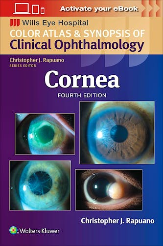
Cornea. Color Atlas and Synopsis of Clinical Ophthalmology. Wills Eye Hospital
Rapuano, C.
4ª Edition June 2024
English
ISBN 9781975214999
Publisher WOLTERS KLUWER
Printed Book
-5%
95,68 €90,90 €VAT included
92,00 €87,40 €VAT not included
Receive it within
2 - 3 weeks
Electronic Book
-5%
80,08 €76,08 €VAT included
77,00 €73,15 €VAT not included
OnLine Access
Immediate
This eBook uses the Bookshelf platform.
What is Bookshelf?
Bookshelf is a tool for managing your eBooks. With online access, you will have a library with all your eBooks. To learn more about Bookshelf, watch an explanatory video .
The eBooks you purchase can be used directly in the browser through the web application Bookshelf Online You can also download them to a Native App to read them anytime and on any device, even when you don't have internet access.
The purchase includes 1 year of Bookshelf Online. The downloaded copy on your device does not expire.
Native Applications
For computers with the following systems:
For mobile devices:
Functionality
The Bookshelf apps offer functionality that goes beyond what printed books provide:
- Online and offline access on mobile or desktop devices
- Bookmarks, highlights, and notes synchronized across all your devices
- Study tools like shared notes and integration with Microsoft OneNote
- Search and navigation of content across your entire Library
- Interactive notebook and text-to-speech functionality
- Online search for additional information by highlighting a word or phrase
Dedication
About the Series
Preface
Acknowledgments
Chapter 1 Conjunctival Infections and Inflammations
Blepharitis and Meibomitis
Chalazion (Internal Hordeolum, Stye)
Bacterial Conjunctivitis (Nongonococcal)
Gonococcal Bacterial Conjunctivitis
Viral Conjunctivitis (Typically Adenovirus)
Chlamydial Conjunctivitis (Adult Inclusion Conjunctivitis)
Trachoma
Molluscum Contagiosum
Ligneous Conjunctivitis
Pediculosis
Parinaud Oculoglandular Syndrome
Ophthalmia Neonatorum
Allergic Conjunctivitis
Atopic Keratoconjunctivitis
Vernal Keratoconjunctivitis
Superior Limbic Keratoconjunctivitis
Floppy Eyelid Syndrome
Toxic and Factitious Keratoconjunctivitis (Keratitis Medicamentosa)
Ocular Rosacea
E-Figures
Chapter 2 Conjunctival Degenerations and Mass Lesions
Pingueculae and Pterygium
Other Conjunctival Degenerations
Amyloidosis
Calcium Concretions
Melanocytic Conjunctival Lesions
Conjunctival Epithelial Melanosis (Racial Melanosis)
Oculodermal Melanosis (Nevus of OTA)
Nevus
Primary Acquired Melanosis
Secondary Acquired Melanosis
Malignant Melanoma
Benign Amelanocytic Conjunctival Lesions
Granulomas
Epibulbar Dermoid
Lipodermoid
Hereditary Benign Intraepithelial Dyskeratosis
Potentially Malignant Amelanocytic Conjunctival Lesions
Squamous Papilloma
Conjunctival Intraepithelial Neoplasia
Squamous Cell Carcinoma
Other Carcinomas
Reactive Lymphoid Hyperplasia and Non-Hodgkin Lymphoma
Cystic Lesions
Primary Conjunctival Cyst
Iatrogenic Cysts
Vascular Lesions
Telangiectasias
Hematologic Disorders
Hemorrhagic Lymphangiectasia
Capillary Hemangioma
Lymphangioma
Kaposi Sarcoma
Sturge–Weber Syndrome (Encephalotrigeminal Angiomatosis)
Carotid–Cavernous Sinus and Dural–Sinus Fistulas
E-Figures
Chapter 3 Anterior Segment Developmental Anomalies
Anomalies of Corneal Size and Shape
Microcornea
Megalocornea
Nanophthalmos
Microphthalmos
Buphthalmos
Congenital Anterior Staphyloma and Keratectasia
Sclerocornea
Cornea Plana
Anterior Segment Dysgeneses
Posterior Embryotoxon
Axenfeld–Rieger Syndrome
Peters Anomaly
Localized Posterior Keratoconus
Aniridia
Iris Coloboma
E-Figures
Chapter 4 Ectatic Conditions of the Cornea
Keratoconus
Pellucid Marginal Degeneration
Keratoglobus
E-Figures
Chapter 5 Corneal Dystrophies
Anterior Corneal Dystrophies
Epithelial Basement Membrane Dystrophy (Anterior Basement Membrane Dystrophy, Map-Dot-Fingerprint Dystrophy, Cogan Microcystic Dystrophy)
Meesmann Corneal Dystrophy (Juvenile Hereditary Epithelial Dystrophy)
Lisch Corneal Dystrophy
Reis–Bücklers and Thiel–Behnke Corneal Dystrophies
Gelatinous Drop–Like Corneal Dystrophy
Stromal Corneal Dystrophies
Granular Corneal Dystrophy, Type I
Lattice Corneal Dystrophy
Granular Corneal Dystrophy, Type II
Macular Corneal Dystrophy
Schnyder Corneal Dystrophy
Posterior Corneal Dystrophies
Fuchs Endothelial Corneal Dystrophy
Posterior Polymorphous Corneal Dystrophy
Congenital Hereditary Endothelial Dystrophy
E-Figures
Chapter 6 Corneal Degenerations and Deposits
Involutional Changes
Corneal Arcus
White Limbal Girdle of Vogt
Crocodile Shagreen
Cornea Farinata
Polymorphic Amyloid Degeneration
Corneal Deposits—Nonpigmented
Band Keratopathy
Salzmann Nodular Degeneration
Other Corneal Degenerations
Spheroidal Degeneration
Lipid Keratopathy
Coats White Ring
Corneal Deposits—Pigmented
Cornea Verticillata (Vortex Keratopathy)
Crystalline Keratopathy
Corneal Iron Deposits
Kayser–Fleischer Ring
Terrien Marginal Degeneration
Iridocorneal Endothelial Syndrome
E-Figures
Chapter 7 Corneal Infections, Inflammations, and Surface Disorders
Bacterial Keratitis
Fungal Keratitis
Acanthamoeba Keratitis
Herpes Simplex Keratitis
Primary Ocular Herpes
Recurrent Ocular Herpes Simplex
Herpes Zoster Keratitis
Interstitial Keratitis (Syphilitic, Nonsyphilitic)
Subepithelial Infiltrates
Superficial Punctate Keratopathy (Punctate Epithelial Erosions)
Thygeson Superficial Punctate Keratopathy
Dry Eye Syndrome (Keratoconjunctivitis Sicca)
Filamentary Keratopathy
Exposure Keratopathy
Neurotrophic Keratopathy
Recurrent Corneal Erosion
Bullous Keratopathy
Acquired Immunodeficiency Syndrome
Contact Lens Complications
Toxic/Allergic Conjunctivitis
Giant Papillary Conjunctivitis
Contact Lens Keratopathy (Contact Lens–Associated Superior Limbic Keratoconjunctivitis and Limbal Stem Cell Deficiency)
Contact Lens Overwear Syndrome
Tight Lens Syndrome
Corneal Warpage
Corneal Neovascularization
Sterile Keratitis
Microbial Keratitis
E-Figures
Chapter 8 Systemic and Immunologic Conditions Affecting the Cornea
Wilson Disease (Hepatolenticular Degeneration)
Vitamin A Deficiency
Cystinosis
Mucopolysaccharidoses and Lipidoses
Collagen Vascular Diseases
Ocular Mucous Membrane Pemphigoid
Stevens–Johnson Syndrome (Erythema Multiforme Major)
Mooren Ulcer
Phlyctenulosis
Staphylococcal Hypersensitivity
Corneal Graft Rejection
E-Figures
Chapter 9 Anterior Sclera and Iris
Episcleritis
Anterior Scleritis (Noninfectious)
Iris Cysts
Iris Pigment Epithelial Cyst
Iris Stromal Cyst
Iris Tumors
Iris Nevus
Malignant Melanoma
Metastatic Tumor
Vascular Tumor
E-Figures
Chapter 10 Surgery and Complications
Cataract Extraction and Intraocular Lens Implantation
Full-Thickness Corneal Transplantation (Penetrating Keratoplasty)
Endothelial Keratoplasty
Anterior Lamellar Keratoplasty
Corneal Biopsy
Superficial Keratectomy
Excimer Laser Phototherapeutic Keratectomy
Conjunctival Flap
Limbal Stem Cell Transplantation
Amniotic Membrane Transplantation
Corneal Perforation
Refractive Surgery
E-Figures
Chapter 11 Trauma
Chemical Burns
Thermal and Electrical Burns
Thermal Burns
Electrical Burns
Ultraviolet Keratopathy (Arc Welder Flash)
Corneal Abrasion
Corneal and Conjunctival Foreign Bodies
Subconjunctival Hemorrhage
Corneoscleral Laceration and Wound Dehiscence
Traumatic Hyphema
Epithelial Downgrowth
Descemet Detachment
E-Figures
Index
Developed at Philadelphia’s world-renowned Wills Eye Hospital, the Color Atlas and Synopsis of Clinical Ophthalmology series covers the most clinically relevant aspects of ophthalmology in a highly visual, easy-to-use format. Vibrant, full-color photos and a consistent outline structure present a succinct, high-yield approach to the seven topics covered by this popular series: Cornea, Retina, Glaucoma, Oculoplastics, Neuro-Ophthalmology, Pediatrics, and Uveitis. This in-depth, focused approach makes each volume an excellent companion to the larger Wills Eye Manual as well as a practical stand-alone reference for students, residents, and practitioners in every area of ophthalmology.
The updated Cornea volume includes:
- Expert guidelines for the differential diagnosis and treatment of cornea diseases seen by the ophthalmic resident, general ophthalmologist, and cornea specialist
- Up-to-date information on infections and complications of corneal surgeries
- More than 450 high-quality photographs of important corneal, anterior segment, and external diseases, many new and updated for this fourth edition
- Revised coverage of the clinical features of key cornea and external eye diseases, diagnostic tests, differential diagnoses, and treatment
- 434 additional full-color photographs and drawings further illustrate pathology and therapeutics described in the text
© 2025 Axón Librería S.L.
2.149.0