
There are no products in the cart.


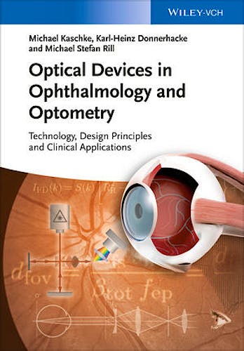
Optical Devices in Ophthalmology and Optometry. Technology, Design Principles and Clinical Applications
Kaschke, M. — Donnerhacke, K.-H. — Rill, M.
1ª Edition January 2014
English
ISBN 9783527648993
Publisher WILEY
Printed Book
-5%
184,96 €175,71 €VAT included
177,85 €168,95 €VAT not included
Receive it within
2 - 3 weeks
Electronic Book
-5%
131,04 €124,49 €VAT included
126,00 €119,70 €VAT not included
OnLine Access
Immediate
This eBook uses the Bookshelf platform.
What is Bookshelf?
Bookshelf is a tool for managing your eBooks. With online access, you will have a library with all your eBooks. To learn more about Bookshelf, watch an explanatory video .
The eBooks you purchase can be used directly in the browser through the web application Bookshelf Online You can also download them to a Native App to read them anytime and on any device, even when you don't have internet access.
The purchase includes 1 year of Bookshelf Online. The downloaded copy on your device does not expire.
Native Applications
For computers with the following systems:
For mobile devices:
Functionality
The Bookshelf apps offer functionality that goes beyond what printed books provide:
- Online and offline access on mobile or desktop devices
- Bookmarks, highlights, and notes synchronized across all your devices
- Study tools like shared notes and integration with Microsoft OneNote
- Search and navigation of content across your entire Library
- Interactive notebook and text-to-speech functionality
- Online search for additional information by highlighting a word or phrase
Description
Medical technology is a fast growing field. This new title gives a comprehensive review of modern optical technologies alongside their clinical deployment. It bridges the technology and clinical domains and will be suitable in both technical and clinical environments. It introduces and develops basic physical methods (in optics, photonics, and metrology) and their applications in the design of optical systems for use in medical technology with a special focus on ophthalmology. Medical applications described in detail demonstrate the advantage of utilizing optical-photonic methods. Exercises and solutions for each chapter help understand and apply basic principles and methods. An associated website run by the authors will include slides to facilitate the teaching/training of this material, and typical images collected by the described methods, eg videos of endoscopy or navigation, OCT,etc.
Table of Contents
Preface
Part One 1
1 Structure and Function 3
- 1.1 Anatomy of the Human Eye 4
- 1.2 Retina: The Optical Sensor 10
- 1.2.1 Retinal Structure 10
- 1.2.2 Functional Areas 12
- 1.3 Recommended Reading 14
- References 14
2 OpticsoftheHumanEye 15
- 2.1 Optical Imaging 15
- 2.1.1 Entrance and Exit Pupils 17
- 2.1.2 Cardinal Points 19
- 2.1.3 Eye Axes 20
- 2.1.4 Accommodation 21
- 2.1.5 Resolution 23
- 2.1.6 Adaption 26
- 2.1.7 Stiles–Crawford Effect 28
- 2.1.8 Depth of Field 29
- 2.1.9 Binocular Vision 30
- 2.1.10 Spectral Properties 32
- 2.2 Schematic Eye Models 33
- 2.2.1 Paraxial Model: The Gullstrand Eye 34
- 2.2.2 FiniteWide-Angle Models 38
- 2.2.3 Applications of Eye Models 44
- 2.3 Color Vision 45
- 2.4 Recommended Reading 47
- References 47
3 Visual Disorders and Major Eye Diseases 49
- 3.1 Refractive Errors 49
- 3.1.1 Axial-Symmetric Ametropia: Myopia and Hyperopia 51
- 3.1.2 Astigmatism 51
- 3.1.3 Notations of Spherocylindric Refraction in Astigmatic Eyes 53
- 3.1.4 Anisometropia 54
- 3.1.5 Distribution of Refractive Errors 54
- 3.1.6 Refractive Errors Caused by Diseases 55
- 3.2 Cataract 56
- 3.3 Glaucoma 57
- 3.4 Age-Related Macular Degeneration 60
- 3.4.1 ARM 60
- 3.4.2 Dry AMD 60
- 3.4.3 Wet AMD 61
- 3.5 Diabetic Retinopathy 64
- 3.6 Retinal Vein Occlusions 65
- 3.7 Infective Eye Diseases 66
- 3.7.1 Trachoma 66
- 3.7.2 Onchocerciasis 67
- 3.8 Major Causes for Visual Impairment 67
- 3.9 Major Causes of Blindness 68
- 3.10 Socio-Economic Impact of Eye Diseases 70
- 3.11 Recommended Reading 72
- Problems to Chapters 1–3 72
- References 76
Part Two 79
4 Introduction to Ophthalmic Diagnosis and Imaging 81
- 4.1 Determination of the Eye’s Refractive Status 82
- 4.2 Visualization, Imaging, and Structural Analysis 82
- 4.3 Determination of the Eye’s Functional Status 85
- 4.3.1 Global Functional Status 85
- 4.3.2 Local Functional Status 86
- 4.4 Light Hazard Protection 86
- References 87
5 Determination of the Refractive Status of the Eye 89
- 5.1 Retinoscopy 91
- 5.1.1 Illumination Beam Path 92
- 5.1.2 Observation Beam Path 93
- 5.1.3 Measurement Procedure 96
- 5.1.4 Accuracy in Retinoscopy 98
- 5.1.5 Applications 99
- 5.2 Automated Objective Refractometers (Autorefractors) 100
- 5.2.1 Common Characteristics of Autorefractors 100
- 5.2.2 Measuring Methods 102
- 5.2.3 Measurement Accuracy and Limitations of Automatic Refractometers 120
- 5.3 Aberrometers 121
- 5.3.1 Fundamentals of Aberrometry 121
- 5.3.2 General Measurement Principles for Aberrometers 126
- 5.3.3 General Remarks on Aberrometry 127
- 5.3.4 Hartmann–Shack Wavefront Aberrometer (Outgoing Light Aberrometer) 127
- 5.3.5 Ingoing Light Aberrometers 131
- 5.3.6 Commercial Aberrometers 133
- 5.4 Wavefront Reconstruction and Wavefront Analysis 133
- 5.4.1 From Wavefront to Refraction (Wavefront Analysis) 135
- 5.4.2 Applications of Wavefront Analysis 140
- 5.5 Excursus: Refractive Correction with Eye Glasses and Contact Lenses 141
- 5.6 Recommended Reading 143
- 5.7 Problems 143
- References 144
6 Optical Visualization, Imaging, and Structural Analysis 147
- 6.1 Medical Magnifying Systems 147
- 6.1.1 Optics of a Single Loupe 148
- 6.1.2 Medical Loupes 149
- 6.2 Surgical Microscopes 151
- 6.2.1 Requirements for Surgical Microscopes 152
- 6.2.2 Functional Principle 154
- 6.2.3 Modular Structure of Surgical Microscopes 160
- 6.2.4 Prospects 176
- 6.3 Reflection Methods for Topographic Measurements 177
- 6.3.1 Keratometer 178
- 6.3.2 Placido Ring Corneal Topographer 187
- 6.4 Slit Lamp 200
- 6.4.1 Functional Principle 201
- 6.4.2 Modular Structure 202
- 6.4.3 Types of Illumination for Various Applications 205
- 6.4.4 Accessories for Other Examinations and Measurements 208
- 6.4.5 Prospects 212
- 6.5 Scanning-Slit Projection Devices 212
- 6.5.1 Lateral Scanning-Slit Projection Techniques 213
- 6.5.2 Scheimpflug Imaging of Rotating-Slit Projections 217
- 6.5.3 Clinical Relevance and Applications 223
- 6.6 Ophthalmoscope 225
- 6.6.1 Functional Principle 226
- 6.6.2 Direct Ophthalmoscope 227
- 6.6.3 Indirect Ophthalmoscope 230
- 6.7 Fundus Camera 236
- 6.7.1 Requirements for a Fundus Camera 237
- 6.7.2 Functional Principle 238
- 6.7.3 Field of View and Magnification 241
- 6.7.4 Wide-Field Imaging 241
- 6.7.5 Color and Monochrome Imaging 241
- 6.7.6 Fluorescence Angiography 242
- 6.7.7 Fundus Autofluorescence 244
- 6.7.8 Stereoscopic Imaging and Analysis 246
- 6.7.9 Equipment Solutions 248
- 6.7.10 Prospects 248
- 6.8 Scanning-Laser Devices 249
- 6.8.1 Confocal Scanning-Laser Ophthalmoscope 250
- 6.8.2 Confocal Scanning-Laser Tomograph 259
- 6.8.3 Scanning-Laser Polarimeter 261
- 6.9 Recommended Reading 267
- 6.10 Problems 267
- References 273
7 Optical Coherence Methods for Three-Dimensional Visualization and Structural Analysis 277
- 7.1 Introduction to Optical Coherence Tomography 278
- 7.2 Development of OCT and LCI as an Example of Modern Medical Technology Innovation 280
- 7.2.1 Academic Research – Conception of OCT (until 1993) 281
- 7.2.2 First Generation of Commercial OCTs (1993–2002) 281
- 7.2.3 Second Generation of OCTs – ZEISS Stratus OCT (2002–2006) 283
- 7.2.4 Third Generation of OCTs – Frequency-Domain OCT (2007–current) 283
- 7.3 Principles of Low-Coherence Interferometry and Optical Coherence Tomography 285
- 7.3.1 Michelson Interferometry with Coherent Light 285
- 7.3.2 Michelson Interferometry with Low-Coherence Light 286
- 7.3.3 Time-Domain OCT 289
- 7.3.4 Frequency-Domain OCT 291
- 7.3.5 Swept-Source OCT 295
- 7.3.6 Overview and Comparison of OCT Systems 297
- 7.4 Elements of OCT Theory 300
- 7.4.1 Theory of Time-Domain OCT – Axial Resolution 301
- 7.4.2 Theory of Frequency-Domain OCT 304
- 7.4.3 Effect of Group Velocity Dispersion in OCT Systems 309
- 7.4.4 Sensitivity and Signal-To-Noise Ratio in TD-OCT and FD-OCT 311
- 7.5 Device Design of OCTs 313
- 7.5.1 Light Sources 313
- 7.5.2 Commercial Systems 315
- 7.6 Ophthalmic Applications of OCT 316
- 7.6.1 Posterior Segment of the Eye 317
- 7.6.2 Anterior Part of the Eye 320
- 7.7 Optical Biometry by Low-Coherence Interferometry 324
- 7.7.1 Dual-Beam Low-Coherence Interferometry 327
- 7.7.2 Applications of Optical Biometry 329
- 7.8 Prospects 334
- 7.9 Recommended Reading 338
- 7.10 Problems 338
- References 341
8 Functional Diagnostics 345
- 8.1 Visual Field Examination 346
- 8.1.1 Physiological Aspects and Functional Principles 346
- 8.1.2 Basic Perimeter Design 351
- 8.1.3 Alternative Perimetric Concepts 357
- 8.1.4 Prospects 362
- 8.2 Metabolic Mapping 363
- 8.2.1 Microcirculation Mapping 363
- 8.2.2 Fluorophore Mapping 366
- 8.2.3 Prospects 367
- 8.3 Recommended Reading 367
- 8.4 Problems 368
- References 368
Part Three 371
9 Laser–Tissue Interaction 373
- 9.1 Absorption 374
- 9.2 Elastic Scattering 375
- 9.2.1 Rayleigh Scattering 376
- 9.2.2 Mie Scattering 376
- 9.3 Optical Properties of Biological Tissue 376
- 9.4 Interaction of Irradiated Biological Tissue 378
- 9.4.1 Photochemical Response 379
- 9.4.2 Photothermal Response 380
- 9.4.3 Photoablation 383
- 9.4.4 Plasma-Induced Ablation and Photodisruption 384
- 9.5 Propagation of Femtosecond Pulses in Transparent Media 391
- 9.5.1 Self-Focusing 392
- 9.5.2 Self-Phase Modulation 392
- 9.5.3 Group Velocity Dispersion 393
- 9.6 Ophthalmic Laser Safety 394
- 9.6.1 Laser Classes 396
- 9.6.2 Safe Use of Ophthalmic Laser Systems 399
- 9.7 Recommended Reading 401
- 9.8 Problems 402
- References 403
10 Laser Systems for Treatment of Eye Diseases and Refractive Errors 405
- 10.1 Laser Systems Based on Photochemical Interactions 406
- 10.1.1 Basics of Photodynamic Therapy 408
- 10.1.2 Technical Equipment Concepts 409
- 10.1.3 Treatment Procedure 411
- 10.1.4 Prospects 411
- 10.2 Laser Systems Based on Photothermal Interactions 412
- 10.2.1 Functional Principle 412
- 10.2.2 Process Parameters 412
- 10.2.3 Treatment Modes 415
- 10.2.4 Technical Equipment Concepts 418
- 10.2.5 Clinical Applications 426
- 10.2.6 Prospects 430
- 10.3 Laser Systems Based on Photoablation 431
- 10.3.1 Basics of Photoablation Treatments 432
- 10.3.2 Technical Equipment Concepts 441
- 10.3.3 Surgical Ablation Techniques 446
- 10.3.4 Prospects 450
- 10.4 Laser Systems Based on Photodisruption with Nanosecond Pulses 450
- 10.4.1 Functional Principle 451
- 10.4.2 Process Parameters 451
- 10.4.3 Technical Equipment Concepts 454
- 10.4.4 Clinical Applications 457
- 10.4.5 Prospects 460
- 10.5 Laser Systems Based on Plasma-Induced Ablation with Femtosecond Pulses 460
- 10.5.1 Functional Principle 460
- 10.5.2 Process Parameters 461
- 10.5.3 Technical Equipment Concepts 463
- 10.5.4 Clinical Applications 466
- 10.5.5 Prospects 472
- 10.6 Recommended Reading 473
- 10.7 Problems 473
- References 476
Appendix A Basics of Optics 481
- A.1 Geometric Optics and Optical Imaging 482
- A.1.1 Refraction and Dispersion 483
- A.1.2 Imaging by Spherical Surfaces 486
- A.1.3 The Ray Tracing Approach to Paraxial Optical Systems 492
- A.1.4 Aperture Stops, Field Stops, and Pupils 496
- A.1.5 Limitations of the Paraxial Beam Approximation 499
- A.1.6 Aberrations 501
- A.1.7 Wavefront Aberration and Image Quality 506
- A.1.8 Classification and Expansion of the Wave Aberration Function 510
- A.1.9 Chromatic Aberration 518
- A.2 Wave Optics 518
- A.2.1 Monochromatic Harmonic Waves 519
- A.2.2 Paraxial Solutions of the Wave Equation 530
- A.2.3 Monochromatic Superposition of Harmonic Waves 535
- A.2.4 Polychromatic Superposition of Waves 537
- A.3 Recommended Reading 543
- A.4 Problems 543
- References 547
Appendix B Basics of Laser Systems 549
- B.1 Einstein’s Two-Level Model of Light–Atom Interaction 550
- B.1.1 Absorption 551
- B.1.2 Spontaneous emission 551
- B.1.3 Stimulated emission 551
- B.1.4 Relation of Einstein Coefficients 552
- B.2 Light Amplification by Stimulated Emission 552
- B.2.1 Conditions for Population Inversion 553
- B.2.2 Multilevel Optical Pumping 555
- B.3 Laser Oscillator 558
- B.3.1 Inversion Threshold 558
- B.3.2 Standing Wave Condition 561
- B.4 The Gaussian Oscillator 563
- B.4.1 Resonator Stability Condition 563
- B.4.2 Divergence 565
- B.4.3 Polarization 566
- B.4.4 Pulsed Laser Operation 567
- B.5 Technical Realization of Laser Sources 571
- B.5.1 Gas Lasers 572
- B.5.2 Semiconductor Lasers 577
- B.5.3 Solid-State Lasers 580
- B.6 Recommended Reading 583
- B.7 Problems 583
- References 588
Appendix C Summary of Used Variables and Abbreviations 591
- C.1 Chapters 1–3 591
- C.2 Chapters 4 and 5 593
- C.3 Chapter 6 594
- C.4 Chapter 7 597
- C.5 Chapter 8 599
- C.6 Chapters 9 and 10 600
- C.7 Appendix A 603
- C.8 Appendix B 605
Index 607
Author Information
Michael Kaschke received his Ph.D. and Dr. sc. nat. degrees both from the University of Jena in 1986 and 1988, respectively, for his research in the field of ultra-short light pulses and spectroscopy. Before joining Carl Zeiss, he was a research scientist at the Max-Born-Institute Berlin, Max-Planck-Institute Göttingen, and invited visiting scholar at IBM Research Center Yorktown Heights, N.Y., working on high-power fs-laser pulses and their matter interaction. He is also a professor for medical technology at the Karlsruhe Institute of Technology, Germany. Michael Kaschke is President and CEO of the Carl Zeiss AG, a technology leader in optics, optoelectronic, and medical technology headquartered in Oberkochen, Germany. He joined Carl Zeiss in 1992 and has held since then several research and management positions in the company predominantly in the medical business.
Karl-Heinz Donnerhacke received his Ph.D. degree from the University of Jena, Germany, in 1976 for his research in the field of high power gas lasers. He spent over 25 years of his career working at Carl Zeiss in laser development and laser application in medicine. During this time, he also joined the University of Jena and the Physical-Technical Institute of the Academy of Sciences as a research scientist. Until his retirement he was Director of R&D in the Ophthalmic Instruments Division of Carl Zeiss for more than 15 years and later Director of R&D for Ophthalmic Diagnostic Instruments at Carl Zeiss Meditec AG. Since 2003, Dr. Donnerhacke has been adjunct professor for ophthalmic technology at the Ernst Abbe University of Applied Sciences Jena, Germany and the Technical University Ilmenau, Germany. Currently, he is also a technical consultant in the field of ophthalmic devices.
Michael Stefan Rill received his Ph.D. degree from the Institute of Applied Physics at the Karlsruhe Institute of Technology, Germany, in 2010 for his research in the field of 3D photonic metamaterials. He subsequently worked as a sales manager at Nanoscribe GmbH in Eggenstein-Leopoldshafen, Germany, and as a scientific assistant to the CEO at Carl Zeiss in Oberkochen, Germany. As of 2013, Dr. Rill holds a position as product manager for cataract surgery systems at Carl Zeiss in Jena, Germany.
© 2025 Axón Librería S.L.
2.149.0