
There are no products in the cart.


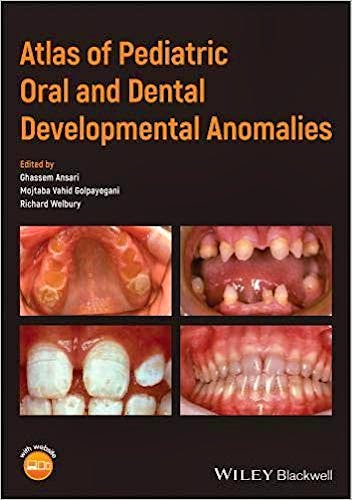
Atlas of Pediatric Oral and Dental Developmental Anomalies
Ansari, G. — Golpayegani, V. — Welbury, R.
1ª Edition February 2019
English
ISBN 9781119380917
Publisher WILEY
Printed Book
-5%
83,19 €79,03 €VAT included
79,99 €75,99 €VAT not included
Receive it within
2 - 3 weeks
Electronic Book
-5%
70,40 €66,88 €VAT included
67,69 €64,31 €VAT not included
OnLine Access
Immediate
This eBook uses the Bookshelf platform.
What is Bookshelf?
Bookshelf is a tool for managing your eBooks. With online access, you will have a library with all your eBooks. To learn more about Bookshelf, watch an explanatory video .
The eBooks you purchase can be used directly in the browser through the web application Bookshelf Online You can also download them to a Native App to read them anytime and on any device, even when you don't have internet access.
The purchase includes 1 year of Bookshelf Online. The downloaded copy on your device does not expire.
Native Applications
For computers with the following systems:
For mobile devices:
Functionality
The Bookshelf apps offer functionality that goes beyond what printed books provide:
- Online and offline access on mobile or desktop devices
- Bookmarks, highlights, and notes synchronized across all your devices
- Study tools like shared notes and integration with Microsoft OneNote
- Search and navigation of content across your entire Library
- Interactive notebook and text-to-speech functionality
- Online search for additional information by highlighting a word or phrase
Preface
About the Companion Website
Chapter I: Oral and Dental Anatomy
The Oral Cavity
1.1 Soft Tissues
1.1.1 The Lips
1.1.2 The Palate
1.1.3 The tongue
1.1.4 The Cheek
1.1.5 Floor of the mouth
1.1.6 The Periodontium
1.2 Hard Tissues
1.2.1 Bone; Maxilla & Mandible
1.2.1.1 The Alveolar Bone
1.2.1.2 Body of Maxilla or Mandible
1.2.1.3 Condyle and TMJ
1.2.2 Teeth
1.2.2.1 Types
Incisors
Canines
Premolars
Molars
1.2.2.2 Dental Anatomy
Crown
Root
Cervical Area
Dental Pulp
1.2.2.3 Occlusion
Chapter II: Histology and Embryology of the tooth and periodontium
2.1 Tooth Histology
2.1.1 Enamel
2.1.1.1 Striae of Retzius
2.1.1.2 Hunter Scherger bands
2.1.1.3 Gnarled Enamel (Spiral enamel)
2.1.1.4 Enamel tufts and Lamella:
2.1.1.5 Enamel surface
2.1.2 Dentine
2.1.2.1 Dentinal Tubules
2.1.2.2 Intra tubular Dentine
2.1.2.3 Inter tubular Dentine
2.1.2.4 Inter globular Dentine
2.1.2.5 Incremental Line
2.1.3 Granular Layer of Tomes
2.1.4 Cementum
2.1.4.1 Cemental connective tissue
2.1.4.2 Fibrous Matrix
2.1.5 Dental Pulp
2.1.6 Periodotium
2.2 Tooth Embryology: Life cycle of the tooth includes the following stages
2.2.1 Initiation (Bud) stage
2.2.2 Proliferation (Cap) stage
2.2.3 Histo-differentiation & Morpho-differentiation (Bell) stage
2.2.4 Apposition and calcification
Chapter III: Epidemiology and Diagnosis of Teeth development disturbances:
3.1 Prevalence and Incidence
3.2 Diagnosis and Classification of Disturbances in Teeth
3.2.1 Source of disturbance
Genetic
Congenital
Acquired
3.2.2 Extent of the involved dentition
Localized
Generalized
3.2.3 Structure involved
Enamel defect
Amelogenesis Imperfecta
Hypoplastic
Hypomature
Hypocalcified
Combined
Dentine defects
Dentinal dysplasia
Dentinogenesis imperfecta
Cementum defects
Hypercementosis
Hypocementosis
Acementosis
Enamel/Dentine/Cementum defect
Odontodysplasia
Odontogenesis Imperfecta
Aplasia (Anodontia)
3.2.4 Teeth Morphology
Invagination
Evagination
Talon Cusp
Gemination
Fusion
Peg lateral
Hutchinson incisors
Mulberry Molars
Double teeth
Odontum
3.2.5 Teeth Size
Macrodontia
Microdontia
Radiation (root shortening)
3.2.6 Teeth Count
3.2.6.1 Hypodontia
Hypodont
Oligodont
Anodont
3.2.6.2 Hyperdontia
Mesiodense
Paramolar
Multiple extra
3.2.7 Color of the teeth
Food and Diet
Vitamins & Minerals
Ions (Fluoride)
Systemic Disease
Cystic fibrosis
High fever
Jundice
Dehydration
Medications
Trauma and infection
Chapter IV: Etiology and Pathology of teeth disturbances
4.1 Genetically originated defects
4.1.1 Disturbances of the teeth Count
4.1.1.1 Reduced in numbers; Missing teeth
Hypodontia
Oligodontia
Anodontia
4.1.1.2 Increased in numbers; Extra teeth
Supernumerary: Conical, Tuberculate, Supplemental Mesiodens
Supernumerary; Para molar
Supernumerary; Natal and Neo natal teeth
4.1.2 Disturbances in proportion and sizes of the teeth
Large Size-Macrodontia
Small Size-Microdontia
Short Roots
4.1.3 Disturbances of the teeth Morphology
4.1.3.1 Dense Invaginatus
4.1.3.2 Dense Evaginatus (Talon Cusp)
4.1.3.3 Peg Shaped laterals
4.1.3.4 Fusion
4.1.3.5 Gemination
4.1.3.6 Dilaceration
4.1.3.7 Concrescence
4.1.3.8 Taurodontism
4.1.3.9 Hutchinson Incisors and mulberry Molars
4.1.3.10 Odontomas
4.1.4 Defects of teeth Structures
4.1.4.1 Enamel Defects
Enamel hypoplasia
Localized Hypoplasia
Generalized Hypoplasia
Amelogenesis Imperfecta
Hypoplastic (Type I)
Hypocalcified (Type II)
Hypomature (Type III)
Mixed – Hypoplastic Hypomature (Type IV)
Congenital Defects
Environmental (acquired) defects
4.1.4.2 Dentine Defects
Dentinal dysplasia
Dentinogenesis imperfect
Dentine Cyst
4.1.4.3 Cementum defects
Hypercementosis
Hypocementosis
Acementosis
4.1.4.4 Enamel/Dentine/Cementum defects
Regional Odontodysplasia
Odontogenesis Imperfecta
Aplasia (Anodontia)
4.2 Congenital Diseases (In Uterus)
4.2.1 Erythroblastosis fetalis
4.2.2 Measles
4.2.3 Rubella
4.2.4 Pneomonia
4.2.5 Purphyria
4.2.6 Syphilis
4.2.7 Dehydration and liquid imbalance
4.3 Acquired (Environmental) Defects
4.3.1 Food and Diet
4.3.2 Vitamins & Minerals
4.3.3 Ions
4.3.4 Diseases and Drugs
infantile jaundice
Liver Disease, Liver transplant
Cystic Fibrosis and Antibiotic therapy
Lead Poisoning
Iron intake
4.3.5 Primary teeth Trauma and tooth infection
4.3.6 Short roots
Chapter V: Eruption Disturbances of Teeth; etiology and diagnosis
5.1 Definition
5.2 Delayed Eruption
5.3 Early Eruption
5.4 Failed exfoliation (primary dentition)
5.5 Early exfoliation /Loss of primary teeth
5.5.1 Localized factors
Trauma
Infection
Neoplasia
5.5.2 Systemic factors
Familial Fibrous Dysplasia (Cherubism)
Acrodynia
Hypophosphatasia
Pseudo Hypophosphatasia
Anomalous dental structures
Systemic diseases
5.6 Failed eruption and Impaction
5.7 Eruption Cyst
5.8 Ectopic Eruption and Transposition
5.9 Labial Frenulum and Lingual Frenulum
5.10 Under eruption-Infra occlusion
5.11 Over eruption
5.12 Palatal and Labial Clefts and Teeth eruption
5-13 Malocclusion
5.13.1 Class I Mal Occlusion
5.13.2 Class II Mal Occlusion
5.13.3 Class III Malocclusion
5.13.4 General Spacing and Diastema formation
5.14 Gingival Overgrowth
Chapter VI: Self evaluation Cases:
Self evaluation Case Answers
References
Written by world-renowned pediatric dentists, this easily accessible, well-illustrated reference covers a wide range of oral and dental developmental anomalies in children and adolescents, and includes rare as well as more common conditions.
Divided into two parts, the first part is dedicated to normal tissue initiation, formation, and development in the orodental region. The second part offers comprehensive pictorial descriptions of each condition and discussions of the treatment options available.
- A useful, quick reference atlas helping students and clinicians diagnose a wide range of oral and dental developmental anomalies in children and adolescents
- Highly illustrated with clinical photographs
- Describes both common and rare conditions, and explores treatment options
Atlas of Pediatric Oral and Dental Developmental Anomalies is an excellent resource for undergraduate dentistry students, postgraduate pediatric dentistry students, and pediatric dental practitioners.
About the Author
Ghassem Ansari DDS, MSc, PhD, FHD, is Professor of Pediatric Dentistry, Dental School, Shahid Beheshti Medical University, Tehran, Iran; and Adjunct Professor of Pediatric Dentistry, European University College, Dental School, Dubai, UAE.
Mojtaba Vahid Golpayegani DDS, MSD, FICD, is Professor of Pediatric Dentistry, Dental School, Shahid Beheshti Medical University, Tehran, Iran.
Professor Richard Welbury MB BS, BDS (Hons), PhD, FDSRCS, FDSRCPS, FRCPCH, HonFFGDP,is Professor of Pediatric Dentistry and Research Lead, School of Dentistry, College of Clinical and Biomedical Sciences, University of Central Lancashire, Preston, UK.
© 2025 Axón Librería S.L.
2.149.0